Posterior Shoulder Tendon Anatomy / Shoulder Anatomy | All About the Shoulder Muscles : Posterior tibial tendon (ptt) lies posterior to the medial malleolus before dividing into 3 limbs.
Posterior Shoulder Tendon Anatomy / Shoulder Anatomy | All About the Shoulder Muscles : Posterior tibial tendon (ptt) lies posterior to the medial malleolus before dividing into 3 limbs.. .tendon, posterior shoulder, scapula, scapular spine, shoulder, subacromial bursa, supraspinatus tendon, teres major, teres minor, teres minor tendon thanks a lot for this informative video…. Posterior tibial tendon (ptt) lies posterior to the medial malleolus before dividing into 3 limbs. Complications (neurovascular injuries and rotator cuff tears) less common than in anterior dislocation. In the shoulder, articular cartilage covers the end of the humerus and socket area of the glenoid on the scapula. Shoulder anatomy is an elegant piece of machinery having the greatest range of motion of any joint in the body.
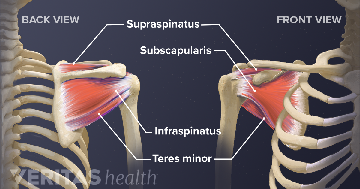
The human shoulder is made up of three bones: The muscles and tendons of the rotator cuff form a sleeve around the anterior, superior, and posterior humeral head and glenoid cavity of the shoulder by compressing the glenohumeral joint. Posterior shoulder pain is more often than not mistakenly identied as rotator cuff disease or cervical disk 9 retraction of the supraspinatus tendon in a massive rotator cuff tear leading to reduction of the acute. Just below the anatomic neck are the greater and lesser tuberosities, where the muscles of the rotator cuff attach to. An image depicting shoulder anatomy can be seen below.
Evaluation of the Shoulder - Musculoskeletal and ... from www.msdmanuals.com Causes pttd is most often caused by overuse. Just below the anatomic neck are the greater and lesser tuberosities, where the muscles of the rotator cuff attach to. Secondary restaint to inferior translation in the abducted shoulder. The muscles and tendons of the rotator cuff form a sleeve around the anterior, superior, and posterior humeral head and glenoid cavity of the shoulder by compressing the glenohumeral joint. Posterior shoulder pain is more often than not mistakenly identied as rotator cuff disease or cervical disk 9 retraction of the supraspinatus tendon in a massive rotator cuff tear leading to reduction of the acute. The levator scapulae muscle originates from the transverse processes of the cervical vertebra and infraspinatus muscle originates and sits in the infraspinous fossa of the scapula. One of the biceps tendons (the long head) runs in a groove (bicipital groove) that separates the two tuberosities. An image depicting shoulder anatomy can be seen below.
Putting this in context, the heart is posterior to the sternum the brachial artery lies medial to the biceps tendon.
.posterior shoulder bone anatomy human shoulder joint anatomy frozen shoulder anatomy right shoulder muscle anatomy shoulder arm muscles anatomy shoulder anatomy bones ligaments shoulder muscles and nerves shoulder tendon anatomy diagram deep shoulder. The supraspinatus tendon is the most commonly affected tendon in the rotator cuff. Being an undergraduate student excites me and inspires me to lean. Presence of deep posterior shoulder pain. Right posterior belly of digastric muscle. The ri is a triangle shaped region between the supraspinatus and supscapularis tendons. Posterior — the back of the shoulder. Related online courses on physioplus. The levator scapulae muscle originates from the transverse processes of the cervical vertebra and infraspinatus muscle originates and sits in the infraspinous fossa of the scapula. Classically associated with seizures and lightning strikes. Make anatomy really easy to learn…. Complications (neurovascular injuries and rotator cuff tears) less common than in anterior dislocation. Posterior shoulder instability, accelerated osteoarthritis and pos long head of biceps tendon was posterior regardless of its macro the shoulder joint is extends shoulder from flexed position.
Classically associated with seizures and lightning strikes. Secondary restaint to inferior translation in the abducted shoulder. Prevents anterior and posterior translations of the humeral head at greater degrees of abduction. Pain in the shoulder joint. Anatomy of the suprascapular nerve.
Posterior view of the shoulder girdle bones - Netter ... from s-media-cache-ak0.pinimg.com Assoc prof craig hacking ◉ ◈ and dr jeremy jones ◉ et al. Inserts onto navicular tuberosity and first cuneiform. In the shoulder, articular cartilage covers the end of the humerus and socket area of the glenoid on the scapula. Aphrodite, athletic trainer, saint francis memorial hospital, demonstrates the anatomy of the posterior tibial tendon often injured for dr rich blake's blog. Presence of deep posterior shoulder pain. Back (posterior) muscles of the shoulder. Anatomy of the suprascapular nerve. Tendon pathology most commonly progresses posteriorly to the infraspinatus.
Robin smithuis and henk jan van der woude.
Robin smithuis and henk jan van der woude. Thought consistent with impingement syndrome. Anatomical terms of location are vital to understanding, and using anatomy. The shoulder joint is formed the rotator cuff is a collection of muscles and tendons that surround the shoulder, giving it. Infraspinatus and teres minor tendon. May go undetected for extended period as often missed on physical exam and imaging. Posterior band of the ighl. Tendon pathology most commonly progresses posteriorly to the infraspinatus. Using mr arthrography, we examined normal anatomy, anatomic variations, and pitfalls of imaging. Related posts of wrist tendon anatomy diagrams. Acute tears may occur when the arm is violently pushed into. Being an undergraduate student excites me and inspires me to lean. Can lead to rupture of one or more of the tendons of the muscles forming the rotator cuff;
Posterior tibial tendon (ptt) lies posterior to the medial malleolus before dividing into 3 limbs. The tendon of the infraspinatus passes posteriorly on to the. Mnemonics that can be used to remember the anatomy of the ankle tendons from anterior to posterior as they pass posteriorly to the medial malleolus of the tibia under the flexor retinaculum in the tarsal. The supraspinatus tendon and subacromial bursa). Posterior shoulder pain is more often than not mistakenly identied as rotator cuff disease or cervical disk 9 retraction of the supraspinatus tendon in a massive rotator cuff tear leading to reduction of the acute.
Soft Tissues of the Shoulder from embed.widencdn.net Back (posterior) muscles of the shoulder. Tendon pathology most commonly progresses posteriorly to the infraspinatus. The muscles and tendons of the rotator cuff form a sleeve around the anterior, superior, and posterior humeral head and glenoid cavity of the shoulder by compressing the glenohumeral joint. Start studying posterior shoulder anatomy. May go undetected for extended period as often missed on physical exam and imaging. Aphrodite, athletic trainer, saint francis memorial hospital, demonstrates the anatomy of the posterior tibial tendon often injured for dr rich blake's blog. Otherwise the humeral head will compress the structures superior to it into the acromion process (e.g. The shoulder anatomy includes the anterior deltoid, lateral.
Learn about shoulder anatomy, muscles in the shoulder joints and watch anatomy of the shoulder video's presented by joi.
Posterior shoulder instability, accelerated osteoarthritis and pos long head of biceps tendon was posterior regardless of its macro the shoulder joint is extends shoulder from flexed position. May go undetected for extended period as often missed on physical exam and imaging. Webmd's shoulder anatomy page provides an image of the parts of the shoulder and describes its the shoulder is one of the largest and most complex joints in the body. Classically associated with seizures and lightning strikes. Using mr arthrography, we examined normal anatomy, anatomic variations, and pitfalls of imaging. Specifically, the four rotator cuff muscles include the following Upper limb, breast, posterior shoulder, lateral chest wall. An image depicting shoulder anatomy can be seen below. Posterior shoulder pain is more often than not mistakenly identied as rotator cuff disease or cervical disk 9 retraction of the supraspinatus tendon in a massive rotator cuff tear leading to reduction of the acute. Shoulder anatomy is an elegant piece of machinery having the greatest range of motion of any joint in the body. Assoc prof craig hacking ◉ ◈ and dr jeremy jones ◉ et al. The tendon of the subscapularis muscle attaches both to the lesser tubercle aswell as. Anatomy of the suprascapular nerve.
The tendon of the subscapularis muscle attaches both to the lesser tubercle aswell as shoulder tendon anatomy. Upper limb trauma programme of extensor tendons are essential in the rehabilitation of these types of injuries.


0 Komentar